| FemaleHealthMadeSimple | |
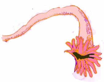
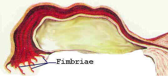
There are two fallopian tubes. The fallopian tubes strech from the upper outside corners of the womb to ovaries. They are tube like structures (like hose pipes). The inner parts are attach to womb and the hollow parts of the tubes connect to and open into the cavity of the womb(uterus). Their outer parts lie near the ovary and the wall of the outer opening end in fingerlike structures called fimbriae. Some of the fimbriae (at least one) is attached to the ovary.
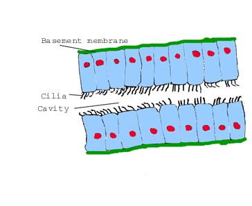
The lining or mucous membrane that lines the cavity of the tube is a special kind of mucous membrane. It consists of a single layer of cells. The cells contain tiny hairlike structures at their surface. These hairlike structures are constantly moving and this movement creatives a sucking power that acts in the direction of the womb's cavity.
Anything in the vicinity of the ovary is sucked into the tube and from there to the womb. This kind of mucous membrane is called a ciliar epithelium.
The next drwawing illustrates the right fallopian tube. The front part of the tube is cut away.
The next drawing illustrates the intact fallopian tube.(Left)
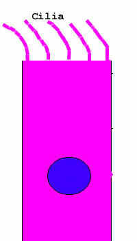
The next drawing show one of the cells of ciliar epithelium.
<
The next illustrate a small segment of the fallopian tube to illustrate the lining (mucous
membrane ) and the ciliar epithelium.
Click the Back button of your browser to go back to the previous
page. ( Usually the left button of the second row).